65 Animal Cell Picture With Labeled Parts
Animal cell diagram without labels images label the cell parts for kids. Sep 21 2018 - Printable animal cell diagram to help you learn the organelles in an animal cell in preparation for your test or quiz.
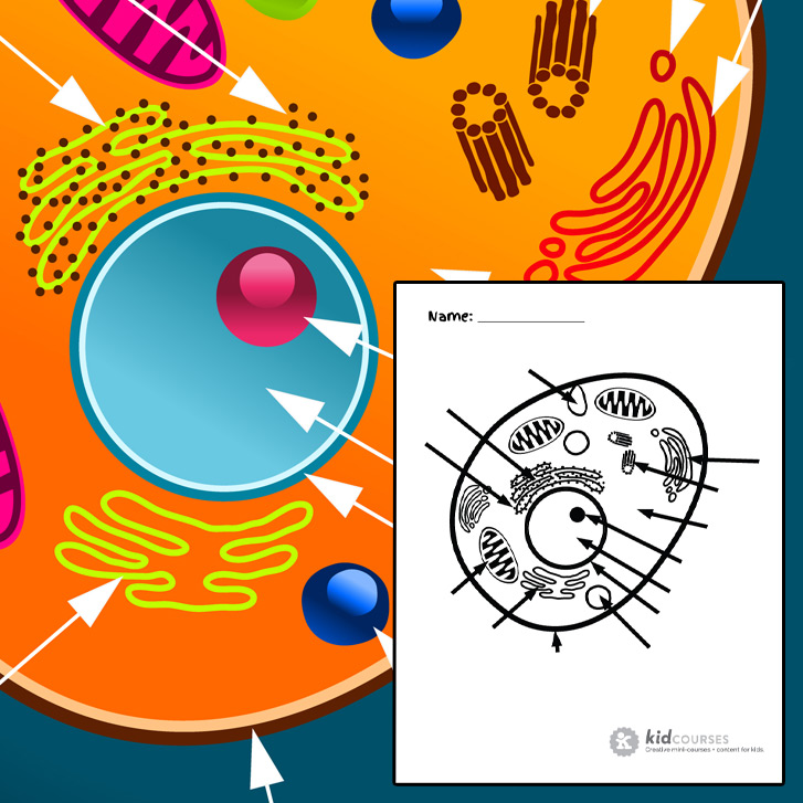
Animal Cell Free Printable To Label Color Kidcourses Com
Find the illustration of the animal cell.

Animal cell picture with labeled parts. A series stack of flattened membrane-bound sacs saccules involved in the storage modification and secretion of proteins glycoproteins and lipids destined to leave the cell extracellular and for use within the cell intracellular. 4 An image of only the outside of the cell. Animal Cell Picture with Labels.
Create a spider map to identify and describe the different parts of the animal cell. Older students can be challenged to identify and label the animal cell parts. Animal Cell Make sure that each part would have a definite function discussed.
Plant cells with their more fixed shape can safely assume that the chromosomes are correctly positioned. 2074 animal cell labeled stock photos vectors and illustrations are available royalty-free. Find a picture and Label the parts of.
Your child will then use the first image to draw the rest of the parts of the cell and label each part. The Golgi apparatus is abundant in secretory cells such as cells of the pancreas. Use the animal cell reference chart as a guide.
In Science go to the anatomy section. Parts of an animal cell. Picture of animal cell labeled.
Eukaryote cell with a nucleus eukaryotic cells vector prokaryotic cells endoplasmic reticulum vector labelled cell microfilaments structure cell animal cells biology prokaryot. 5 Labels for the animal cell its parts and some blank labels in case you wanted to teach more than 8 parts. Identify the different parts of the animal cell and type them into the title boxes.
How to draw a Animal Cell easy and step by step. A membrane-bound body that forms by. Get The Markers HERE httpsamznto37ZBdoN.
Younger students can use the animal cell worksheets as coloring pages. Plant and animal cell worksheet plant cell structure and function worksheet and plant cell diagram without labels are three of. Your child will use the first image as reference or just draw it and label it all by heart.
3 An image of the animal cell with only the nucleus in the center for reference. Download Animal cell structure stock photos. Labeled animal cell with Truffy Nov 22 0300 PM 1.
Affordable and search from millions of royalty free images photos and vectors. The guy who sent this email undoubtedly sniffs his own farts. Controls passage of materials in and out of the cell cytoplasm everything inside of the cell membrane except for the nucleus light yellow nucleus control center of the cell.
Draw this Animal Cell by following this drawing lesson. Cell membrane surrounds the internal cell parts. See animal cell labeled stock video clips.
In eukaryotic cells the cytoplasm includes all of the material inside the cell and outside of the. This is a great image for quizzing. 5th grade science and biology.
Each cell should have one part of the diagram colored a different color than the rest matching the title box. Contains DNA light pink. Plant cell Cytoplasm - is a thick solution that fills each cell and is enclosed by the cell membrane.
It is mainly composed of water salts and proteins. Label parts and thousands of other science skills. Since animal cells are softer than plant cells centrioles are required to ensure the chromosomes are in the proper location when the cell divides.
Talking related with Labeled Plant Cell Parts Worksheet scroll down to see particular similar images to complete your ideas. Improve your science knowledge with free questions in Animal cell diagrams. Nov 8 2013 - Animal Cell Model Labeled picture of animal cell labeled.
Find Structure Animal Cell Labeled Parts Biology stock images in HD and millions of other royalty-free stock photos illustrations and vectors in the Shutterstock collection. Printable Animal Cell Diagram Labeled Unlabeled And Blank In dp2 if you quicklook a chaptered movie you see the chapters with thumbnails listed vertically in a left pane of the quicklook window doesnt work quite well yet. Thousands of new high-quality pictures added every day.
90 Animal Cell Vacuole Diagram
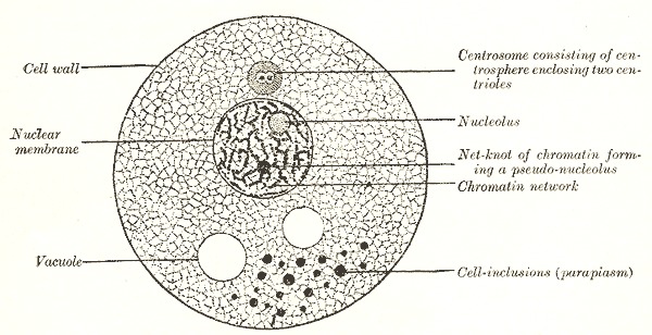
Nucleus usually found near the center of an animal cell and along the edge of a plant cell holds the organisms genetic information and directs most all of the activities in the cell. The vacuole is a type of organelle present in eukaryotic cells.
:max_bytes(150000):strip_icc()/animal_cell-56c765663df78cfb3788382b-5c2e861046e0fb000142aa47.jpg)
Animal Cells And The Membrane Bound Nucleus
Animal cell diagram detailing the various organelles Though this animal cell diagram is not representative of any one particular type of cell it provides insight into the primary organelles and the intricate internal structure of.
Animal cell vacuole diagram. The structure of the plant cell is. In plant cells collects radiant light energy from the sun and uses it to convert carbon dioxide CO2 and water H2O into a sugar glucose. Plant Cell Diagram Vacuole.
Everyone in the town has something to do with steel widget making and the entire town is designed to build and export widgets come in all shaped and. Plant vs animal cells diagram. In yeast cells the vacuole is.
It is a sac surrounded by a single membrane called a tonoplast. The main function of vacuoles in animal cells is to isolate and remove waste products from the other organelles and the cytoplasm. Although animal cells contain vacuoles they do not contain large central vacuoles.
A vacuole is usually found in all plant and fungal cells as well as some cells of protists animals and bacteria. Vacuole Function in Animal Cells. Plant vacuole contains cell sap.
The vacuole has an important structural function as well. Vacuoles serve many functions such as supporting the cell wall in plant cells. As the central vacuole shrinks it leaves the cell wall unsupported.
Animal cell diagram labeled vacuole. Animal Plant Nucleus Cytoplasm Cell membrane Ribosome Mitochondria Vacuole Cell wall Chloroplast. The various cell organelles present in an animal cell are clearly marked in the animal cell diagram provided below.
The membrane holds fluid called cell sap which is composed of water and other substances. Vacuoles are permanent structure in plant cell. Cell membrane cell wall chloroplast cytoplasm mitochondria nucleus ribosome vacuole Cell City Analogy In a faraway city called Grant City the main export and production product is the steel widget.
A bacteria diagram clearly enables us to learn extra approximately this single cell organisms that have neither. Vacuoles are generally occurs in center of plant cell. Animal vacuole contains fluid food or metabolic waste in animal cell.
The vacuole can be a water tower because a water tower stores water and the vacuole also stores water along with a couple of other things like nutrients. Vacuoles generally have acidic pH values. A vacuole is an organelle in cells which functions to hold various solutions or materials.
The food vacuole contains digestive enzymes with the help of which nutrients are digested. Animal cells usually have an irregular shape and plant cells usually have a regular shape. Lysosome Answers may vary Possible Venn diagram Pl antAnimal Cell Venn Diagram Pl ant Cel l s Ani mal Cell s B oth T ypes of Cell s - Have a rigid cell wall outs ide of the cell membrane that provides s tructure and s upport - Have chloroplast.
Cellcell wall a cell p label. The ribosomes can be a restaurant. Vacuoles can be temporary structure in animal cell.
Nucleus Ribosomes Rough Endoplasmic Reticulum Smooth Endoplasmic Reticulum Mitochondria Golgi Apparatus Cytoplasm Vacuole Cell Membrane. It is mainly made up of water and protein material. Hall controls everything in the city and its just like the nucleus because the nucleus controls want happens in the cell.
RETURN to CELL DIAGRAM. ANIMAL CELLSUse the words in the word box. A vacuole is a membrane bound structure found in the cytoplasmic matrix of a cell.
While animal cells may have many tiny vacuoles a plant cell usually has a single large vacuole which serves as a storage tank for food water waste products and other materials. In animal cells vacuoles tend to play a lesser role. Animal cell contains one or many Vacuoles which are smaller in size as compared to plant vacuoles.
Vacuoles are distributed all over the animal cell. Animal cells have one or more small vacuoles while plant cells have a large central. The cell membrane is the outer most part of the cell which encloses all the other cell organelles.
28 Animal Cell Diagram With All Organelles
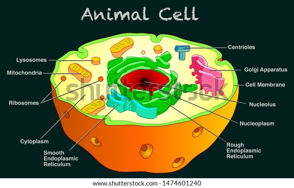
A typical animal cell consists of the following parts. He explains each organelles function including the nucleus nucleolus nuclear envelope nuclear po.
1
Start studying Animal cell organelle.

Animal cell diagram with all organelles. Parts of Animal Cells. Learn vocabulary terms and more with flashcards games and other study tools. 5 Rough endoplasmic reticulum RER.
Animal Cell - Science Quiz. Animal cells are packed with amazingly specialized structures. The nucleus contains all of the genetic material in a cell.
The cell membrane is the outer most part of the cell which encloses all the other cell organelles. Though this animal cell diagram is not representative of any one particular type of cell it provides insight into the primary organelles and the intricate internal structure of most animal cells. Each cell can be thought of as a large factory with many departments like manufacturing packaging shipping and accounting.
A Cell membrane A Nucleus The Cytoplasm The various organelles. The structure of the Golgi Complex is pleomorphic. There are various organelles present within the cell and are classified into three categories based on the presence or absence of membrane.
Animal cell diagram and functions of organelles. Furthermore it is easy to distinguish between a plant and animal cell diagram just by inspecting the presence or absence of a cell wall. However it typically exists in three forms ie.
Outer membrane of cell that controls movement in and out of the cell Double layer. This animal cell diagram doesnt represent any particular Animal cell it provides insight into the primary organelles and the internal structure of the most Animal cell. Differences in cellular structure of prokaryotes and eukaryotes include the presence of mitochondria and chloroplasts the cell wall and the structure of chromosomal DNA.
The various cell organelles present in an animal cell are clearly marked in the animal cell diagram provided below. The membrane is selectively permeable and allows only certain molecules. Its the cells brain employing chromosomes to instruct other parts of the cell.
Animal cell diagram detailing the various organelles. Animal Cell Definition Structure Parts Functions And Diagram The animal cell has 13 different types of organelles 1 with specialized functions. The cell membrane is a double-layered membrane made up of phospholipids that surrounds the entire cell.
The mitochondria are the cells powerplants combining chemicals from our food with oxygen to create energy for the cell. An animal cell diagram is shown above clearly shows the various cell organelles present in the animal cell. Eukaryotic cells contain membrane-bound organelles such as the nucleus while prokaryotic cells do not.
8 Smooth endoplasmic reticulum SER. A tour of the animal cell by Biology Professor Dr. It is mainly made up of water and protein material.
Cell membrane nucleus nucleolus nuclear membrane cytoplasm endoplasmic reticulum Golgi apparatus ribosomes mitochondria centrioles cytoskeleton vacuoles and vesicles. All cell organelles are marked clearly in the diagram. Animal cell organelles labeled diagram.
One vital part of an animal cell is the nucleus. An animal cell is the basic unit of any living animal. Diagram showing the structure of a cell membrane.
A group of cells forms tissue various tissues forms an organ and different organs make up the body. A labeled diagram of the animal cell and its organelles there are two types of cells prokaryotic and eucaryotic. Its a very simple way to distinguish between animal and plant cell diagrams just by.
Animal cell diagram detailing the various organelles Though this animal cell diagram is not representative of any one particular type of cell it provides insight into the primary organelles and the intricate internal structure of most animal cells. Main parts of an animal cell. All the cells found in any living animal are made up of similar components and organelles and are eukaryotic cells.
The various cell organelles present in an animal cell are clearly marked in the animal cell diagram provided below. Parts of the animal cell structure. The cell organelles found in the animal cell are plasma membrane centriole peroxisome lysosome ribosomes mitochondria endoplasmic reticulum cytoplasm nucleus nucleolus nuclear envelope and golgi apparatus.
Listed below are the Cell Organelles of an animal cell along with their functions. The well labelled diagram of an animal cell consists of all the organelles and the structural components of an animal cell. The cell membrane controls the influx of the nutrients and minerals in and out of the cell.
The Golgi Apparatus is the cell organelle mostly present in eukaryotic cells which is responsible for the packaging of macromolecules into vesicles so that they can be sent out to their site of action. Cisternae vesicles and tubules. 11 Cytosol Its not an organelle.
Its the fluid that contains the organelles.
83 Animal Diagram
Rabbit dragon snake horse goatram monkey rooster dog pig rat ox and tiger. Ribosome small organelles composed of rna rich cytoplasmic granules that are sites of protein synthesis.
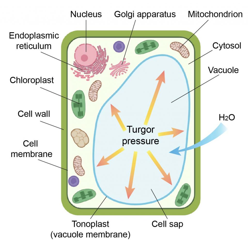
Animal Cell Diagram Cross Section Annotated Stock Vector Royalty Free 1474601240
Forest Animals in English A Label Me.

Animal diagram. The diagram like the one above will include labels of the major parts of an animal cell including the cell membrane nucleus ribosomes mitochondria vesicles and cytosol. The animal cell diagram is widely asked in Class 10 and 12 examinations and is beneficial to understand the structure and functions of an animal. Animal cells usually have an irregular shape and plant cells usually have a regular shape.
Jan 7 2020 - Explore sunny bests board animal diagrams followed by 298 people on Pinterest. Animal Farm by George Orwell is a dystopian vision of society based on the early years of communist Russia. Here is a picture gallery about dyson dc25 animal parts diagram complete with the description of the image please find the image you need.
Animal cell diagram and functions. Boxer the horse worked long hours pulling stone for the windmill. With the help of this diagram of an animal cell quiz online youll have the chance to develop your understanding of the different structures and organelles in an animal cell and better relate them to their corresponding functions.
Ereplacementparts pertaining to Dyson Dc25 Animal Parts Diagram image size 620 X 806 px and to view image details please click the image. Animal Class Diagram UML Use Createlys easy online diagram editor to edit this diagram collaborate with others and export results to multiple image formats. This is such a cute animal science activity for early grades.
See more ideas about drawings animal drawings animal sketches. When you spin the wheel the 12 animals of the Chinese zodiac are displayed the picture name of the animal and the years that it represents one at a time. This ancestor diversified over time into several descendent subgroups which are represented as internal.
About a week later all their progress was destroyed. Here at Animal Corner we have compiled some of the most comprehensive diagrams and descriptions of all aspects of Animal anatomies so. The cells of animals are the basic structural units for the wide variety of life we see in the animal kingdom.
An animal cell diagram is a great way to learn and understand the many functions of an animal cell. So were sharing a fun and simple animal science lesson for keeping up with animal diets and habitats a Venn diagram animal sorting activity. Gray Fox Skull Diagram and Labeling.
Though this animal cell diagram is not representative of any one particular type of cell it provides insight into the primary organelles and the intricate internal structure of most animal cells. Hummingbird Skull Diagram and Labeling. 163968910 stock photos online.
Llama Thoracic Abdominal Organs Left View Llama Thoracic Abdominal Organs Right View Llama Venipuncture - Drawing Blood in the Llama. Dyson Dc25 Parts List And Diagram. The root of the current tree connects the organisms featured in this tree to their containing group and the rest of the Tree of Life.
Plus it can work as a science center activity you can reuse. Animal Anatomy Anatomy is the branch of biology concerned with the study of the structure of organisms and their parts. Napoleon sniffed pig hoof prints and said that Snowball destroyed the windmill.
Label a diagram of a flamingo. We were unable to load the diagram. It is an allegory filled with elements of what can happen in the wake of a popular revolution.
A brief explanation of the different parts of an animal cell along with a well-labelled diagram is mentioned below for reference. Horse External Anatomy Horse Digestive Tract. Printout Label the fox squirrel deer antler bear claw raccoon hedgehog mouse and worm in English.
Frog Life Cycle Diagram Label a diagram of the frogs life cycle. The most important structures of plant and animal cells are shown in the diagrams below which provide a clear illustration of how much these cells have in common. The animals started building the windmill for electricity.
Furthermore it is easy to distinguish between a plant and animal cell diagram just by inspecting the presence or absence of a cell wall. The basal branching point in the tree represents the ancestor of the other groups in the tree. This tree diagram shows the relationships between several groups of organisms.
Download 7718 Animal Diagram Stock Illustrations Vectors Clipart for FREE or amazingly low rates. Tap diagram to zoom and pan. The latter is the space that occupies maximum part of the cell and where the cell organelles are present.
New users enjoy 60 OFF. Like many dystopias the societys goal was to build a utopia where its members live in harmony but these ideals quickly transformed into something darker. The significant differences between plant and animal cells are also shown and the diagrams are.
62 3 Organelles Found In Plant And Animal Cells
Mitochondria Cell Wall Cytoplasm. Just as it sounds this is the skeleton or structure for cell support.

Animal Cell Definition Structure Parts Functions And Diagram
Membrane-bound organelles such as the nucleus mitochondria endoplasmic reticulum golgi apparatus lysosomes and peroxisomes.
3 organelles found in plant and animal cells. The genetic information is. Cells are the fundamental units of life on Earth and they are the building blocks that make up all other living things. Structural support of cells.
Controls the movement of substances into and out. They both contain membrane-bound organelles such as the nucleus mitochondria endoplasmic reticulum golgi apparatus lysosomes and peroxisomes. 7 rows Part Function Found in.
808 views There was an error loading more items. Cell Wall - Cellulose-containing cell wall that gives plant cells their shape. This stores several pigments ions enzymes and organic and inorganic substances.
It also has a great role in osmoregulation. Large Central Vacuole - although. Facilitates the movement of organelles.
Processes packages and distributes proteins to other organelles for export. This provides extra strength and support for the plant cell so it doesnt burst when gaining water by endosmosis. Assignment 3 Cells and their organelles Cell membrane Cell wall Plant cell Cytoplasm Eukaryotic Nucleus Ribosome Sooth ER Protist Archae Centrosomes Cytosol Animal cell Bacteria Central vacuole Microscope Prokaryotic Rough ER Mitochondria Nudear pore Chloroplast Lysosomes Cytoskeleton Diploid Haploid Fill in the blanks with a word or.
Differences Between Plant And Animal Cells You can observe in these plant and animal cell diagrams. Contains and protects the genetic material in a cell. Plant cells have a cell wall chloroplasts.
An organelle that stores water and food in plant and animal cells. Which set of organelles are found in all three types of cells. Chloroplast - The organelle where photosynthesis occurs.
Animal cells are also known as eukaryotes. Organelles found only in animal cells and their jobs. The cell wall of plants is made of.
These differences can be observed under the electron microscope. Simply so what are the main differences between plant and animal cells. Animal cells contain these cylindrical structures that organize the assembly of microtubules during cell division.
This assists a cell when it is dividing in a process called mitosis and meiosis. This is where photosynthesis takes place. In case of animal cells plastids are not found and are completely absent.
Contains the genes chromatin. This is additional support for the structure of the cytoskeleton. Chromoplasts chloroplasts and leucoplasts.
An organelle where energy production occurs. Animals cells do not have a cell wall unlike plant cells. It confers shape and rigidity.
Eukaryotic animal cell diagram 3 plant cell differences. 14 rows The cytoplasm is present both in plant and animal cells. Plastids are found in plant cell and they are of three types.
Every cell contains a set of organelles. Subcellular structures that are specially adapted to carry out the necessary functions of life. A jellylike material that makes up the inside of a cell.
An organelle that contains the genetic material controlling the activities of the cell. They are jelly-like substances. Three organelles that are present in plant cells but not present in animal cells are.
Some organelles including the nucleus mitochondria and endoplasmic reticulum are found in virtually all eukaryotic cells. Structurally plant and animal cells are very similar because they are both eukaryotic cells. Answered by Lifeeasy Authors.
Both also contain similar membranes cytosol and cytoskeletal elements. Protects the cell from osmotic swelling. Some of the key eukaryotic organelles and their functions are.
Ribosome Cell Membrane Vacuole.
